Science
in Christian Perspective
Science
in Christian PerspectiveScience
in Christian Perspective
Frank Allen, M.A., Ph.D., LL.D, F.R.S.C.
Professor
Emeritus in Physics
University of Manitoba, Winnipeg, Canada
From: JASA 1 #2 (May 1949): 9-20.
"Sturmius, says Paley, held that the examination of the eye was a cure for atheism'. Yet Helmholtz, who knew incomparably more about the eye than half a dozen Sturms, describes it as an instrument that a scientific optician would be ashamed to make; and Helinholtz was no atheist."
This quotation from Professor Ward's Gifford Lectures1 is a reference to the celebrated popular lecture2 of Helmholtz on vision. In view of the commanding influence on visual science which he has exerted for over ninety years since the beginning of the publication of his Handbuch der Physiologischen Optik (1856-66), his views on the characteristics of the eye as an optical instrument carry great weight. The importance of this subject from several standpoints has led the writer to assemble the latest measurements on the defects of the eye, as well as all the statements of scientific men of unquestioned authority in this field of enquiry that he could find.
First of all, the words of Holmholtz himself are given so that there may be no doubt as to what he did say.
"Now it is not too much to say that if an optician wanted to sell me an instrument which had all these defects, I should think myself quite justified in blaming his carelessness in the strongest terms, and giving him back his instrument. Of course, I shall not do this with my eyes, and shall be only too glad to keep them as long as I can--defects and all. Still, the fact that, however bad they may be, I can get no others, does not at all diminish their defects, so long as I can maintain the narrow but indisputable position of a critic on purely optical grounds .... All these imperfections would be exceedingly troublesome in an artificial camera and in the photographic picture it produced. But they are not so in the eye--so little, indeed, that it was difficult to discover some of these .... The chief reason (for not observing the defects) is that we are continually moving the eye, and also that the imperfections almost always affect those parts of the field to which we are not at the moment directing our attention ....For the eye has every possible defect that can be found in an optical instrument, and even some which arc peculiar to itself but they are all so counteracted, that the inexactness of the image which results from their presence very little exceeds, under ordinary circumstances of illumination, the limits which are set to the delicacy of sensation by the dimensions of the retinal cones .... The adaptation of the eye to its function is, therefore, most complete, and is seen in the very limits which are set to its defects .... The defects which result from the inexactness of vision and the smaller number of cones in the greater part of the retina are compensated by the rapidity with which we can turn the eye to one point after another of the field of vision, and it is this rapidity of movement which really constitutes the chief advantage of th eye over other optical instruments."
The defects of the eye, which are set forth so prominently in this discussion, should be reviewed in the light of the most recent knowledge of visual optics which has been assembled both by Duke-Elder3 in his exhaustive treatise on ophthalmology, and also by Hartridge4 in his recent investigation of the visual perception of fine detail. For no optical instrument has been examined so minutely, critically and accurately as the eye, by men of the highest genius such as Helmholtz, Gullstrand and Tscherning.
In the optical system of an instrument, such as the microscope or a telescope, the surfaces of the lenses should be perfectly spherical and they should be centered upon a common optical axis. The mathematical theory of such a dioptric system, as originally developed by Gauss, demands, in addition, that it should be of small aperture, not more than 100, so that the rays of light are limited to the axial regions, of homogeneous media, and that monochromatic light should meet the refracting surfaces nearly at right angles. It is with such an ideal system that all practical optical instruments are compared.
Because of the nature of light and its mode of propagation, as well as the nature of the refracting media which form the image of an object, all optical systems are subject to many aberrations. Those defects must in some way be overcome or minimized or the image will be more or less imperfect. The method of optical correction is to use combinations of several lenses of different refractive powers and curvatures. In the microscope, an achromatic object glass, which forms the image, is composed of as many as six lenses, and an apochromatic object glass may have ten. These lenses must be made of carefully selected glass with accurately figured surfaces, and adjusted to each other with extreme precision. The focal length is quite small. So perfect can the image be made, that magnifications of more than 1500 diameters are possible. They operate at a fixed distance from the object under observation, and the field of view is extremely small. In addition to the object glass there is the eyepiece with which the image is magnified and viewed. All the lenses are immersed in air so that the numerous surfaces have abrupt discontinuity with that medium. The telescope has fewer lenses, but the focal length is relatively large which minimizes or eliminates the most troublesome defects.
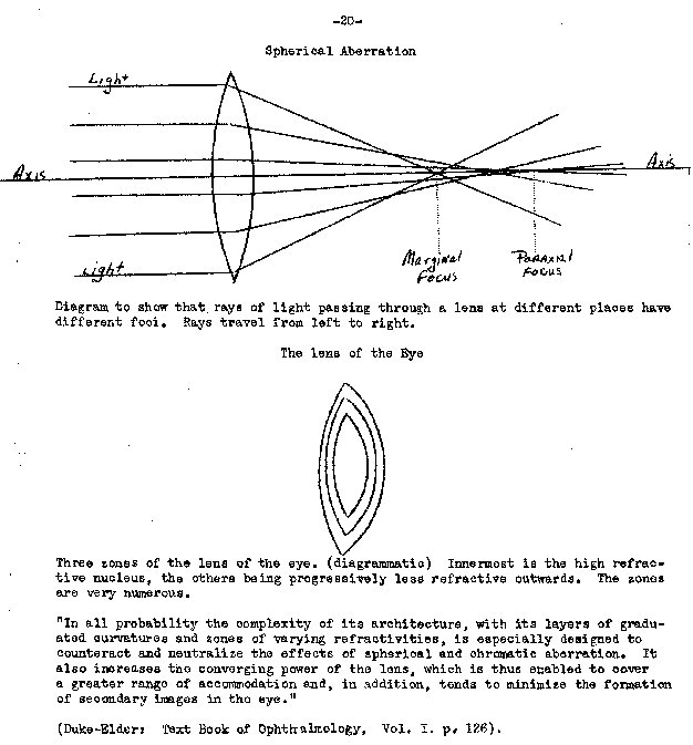
Compared with such complicated optical instruction, the human eye is rudimentary. The refracting system consists of the cornea, the aqueous humor, the lens and the vitreous humor or body. The lens, therefore, is immersed in the humors and not in air, which greatly minimizes the optical discontinuities. The indices of refraction of the two humors are practically the same (1.336), which differs but little from that of water (1.334). The lens is extraordinary in constitution since it consists of many layers or zones which vary in refractive index from 1.386 at the surfaces to 1.406 at the canter. The cornea and aqueous humor taken together, form the major refracting medium of the eye, and the lens, by its power of accomodation, operates as the fine adjusting mechanism. The cornea-aqueous combination is about 2.5 times greater in refracting power than the lens, which is due to the relatively great difference in refractive index between the air (1.00) and the cornea (1.376), as compared with (refractive index) between the humors (1.336) and the surface of the lens (1.386). The rays of light are first refracted at the cornea and aqueous humor, second by the anterior zones of the lens, then by the core, and further by the posterior zones. In. consequence of its structure, the lens has a greater refracting power if it were homogeneous with a refractive index equal to that of the core. Besides increasing the refractive power considerably, this arrangement diminishes the spherical aberration.
The fact of its decontration constitutes the main feature of the optical system of the eye. Strictly the cornea has no axis of symmetry, though the deviation is extremely small. The centering of the cornea and the two surfaces of the lens is never exact, nor are the various zones of the lens concentric with one another, and the lens itself is not concentrically placed upon the optic axis. The visual axis, which touches the fovea, makes an angle of about 5 with the optic axis upon which all refracting surfaces should ideally be centered. Vision therefore occurs obliquely through the system. "It is," remarks Duke-Elder, "as if one of the lenses of an optical instrument had slipped a little out of place and then when we looked through the instrument we tilted it very slightly. The deviations, however, are usually so small as to be functionally negligible, and they tend to some extent to minimize the aberrations of the eye."
It would seem, therefore, that the construction of the eye slightly violates all the conventions that apply to optical instruments. But the final result, nevertheless, is the formation of an improved image, as free from perceptible defects as those produced by the finest artificial systems.
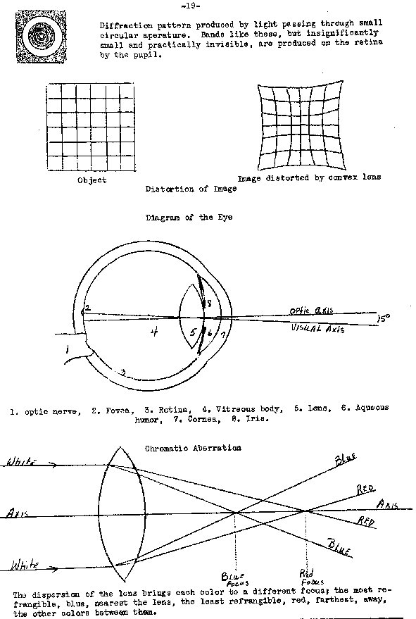
The defects of dioptric systems, whether of glass or of the eye, comprise two main classes: first, aberrations originating from the composite nature of white light, termed chromatic aberration, and those arising from the structure of light and the manner of its propagation, called diffraction; second, monochromatic aberrations, or defects occuring with rays of light of a single wavelength; spherical aberration, the sine condition, refraction of eccentric rays, curvature and distortion of the image, and depth of focus, which is not a defect but a property of a lens.
The defects of the first group depend on the nature of light; those of the second group on the structure of the optical instrument.
The discovery of the spectrum by Newton (1664) established the composite nature of white light and the different refrangibilities of the colors. From these discoveries he erroneously concluded that an achromatic combination of lenses was impossible to devise. Apparently he gave no thought to the construction of the eye. As no chromatic aberration had ever been observed in it, Euler (1750), convinced that it was achromatic, stimulated the eminent optician Dollend (1758) to construct achromatic systems of lenses, such as telescopes, in which he was highly successful. He also discovered the chromatic aberration of the eye.
Since white light is a mixture of many different wavelengths and as a lens has the nature of a series of prisms, the real image of a disc of white light formed by a convex lens consists of a superimposed series of images of all colors of the spectrum focused at slightly different distances from the lens. The violet image, formed by the most refrangible rays, is nearest the lens, and the red image the most remote. The chromatic difference of focus is very small, being from red to violet, about 0.47 mm. (Hartridge). Since the eye accommodates itself to the brightest yellow-green rays, the difference of focus is normally about half the amount for violet in front of the retina, and half for red, as it were, behind it. The separate color images or aberration discs are also of different sizes, termed the chromatic difference in magnification, varying, according to Hartridge, for the usual entrance pupil of 4 mm. diameter for the eye, from 0.0216 mm. in diameter for orange light to 0.0588 mm. for blue, the latter being 2.7 times the former. In ordinary vision these defects are extremely small, and since the blue image completely overlaps the red, it neutralizes that color into white leaving a narrow blue border which, because of its low luminosity, is practically invisible. The whole effect of chromatic difference in magnification tends to counteract the chromatic difference of focus. In the eye the effects of chromatic aberration are small; and with a 2 mm. diameter pupil, as Duke-Elder remarks, 70 per cent of the light falls on a retinal area of 0.005 mm. diameter. The remaining light is more widely diffused and normally is therefore unnoticed, so that images are free from colored borders, as common observation shows.
Diffraction is due to the spreading of the waves of light at the edges of the wave front. The effect is the opposite of chromatic aberration since it is greatest with the longest waves, the red, and least with the shortest, the violet. It follows from the nature of light that a point of white light is always brought to a focus as a small blurred disc (Airy's disc) of light and dark bands with a bright spot in the center. No point image can therefore be formed but only a diffraction pattern, which varies in dimensions directly as the focal length of the system and the wavelength of light, and inversely as the aperture, or area of the pupil, through which the light passes. Diffraction is inherent in the nature of light and its propagation and cannot be overcome. In optical instruments, such as the microscope, its effects are not troublesome except at very high magnifications about 2000 diameters, when the image itself becomes a complicated set of diffraction patterns.
The central bright spot of the diffraction pattern of a point source receives
about 84 per cent of the incident light; the first ring 1/57th and the second
1/240th of the intensity of the central area. At a distance of only 0.02 nun.
from the center, the light is of too low an intensity to be seen. Since the eye
has a very short focal length of 22.78 mm., the diffraction effects with a pupil
4 mm. in diameter are correspondingly small.
Comparison of the figures for
chromatic aberration and diffraction shows that they have the same order of
magnitude, and are much too slight to be observed in the ordinary usage of the
eyes. It is to be noted that the effects of chromatic aberration increase as the
pupil widens; but as this condition changes act together to leave the actual
definition of the image practically unchanged with alterations of the pupil. As
pupillary areas are governed by the illumination, the definition of the image is
therefore the same with all intensities of light.
Those are the
characteristics of the images of point sources. But "the extended source",
according to Hartridgc, "differs from the point source in an important respect,
namely, that its image is largely composed of white light, and that color
fringes are only found near its margins. Each of the fringes for a 4 mm. pupil
is about 0.01 mm. wide. Only parts near the edge are strongly colored, and that
as the distance from the edge increased the yellow fringes become progressively
whiter and the blue fringe progressivly blacker, until the yellow fringe merges
imperceptibly with the white interior of the image, and the blue fringe merges
similarly with the black background."
The mathematical theory of optical
systems of lenses demands a very small aperture so that the rays are limited to
the axial regions. In optical instruments an aperture of 100 is considered to be
the maximum compatible with efficiency. It is with this aperture that such
instruments are designed to eliminate chromatic aberration and other defects.
But the pupil of the eye is rarely less than 4 mm. in diameter which corresponds
to an aperture at the cornea of 200. With a double size of aperture, therefore,
the eye is equally free from aberrations, while optical instruments with such
wide apertures would probably be useless.
Spherical aberration
In a biconvex
spherical lens the peripheral or marginal rays are refracted more than the
axial, so that the former come to a focus nearer the lens than the latter. This
effect is called spherical aberration. It can be eliminated by grinding the lens
so that its curvature decreases from the center to the periphery. Such lenses
are termed aplanatic. Spherical aberration is diminished by making the curvature
of the anterior surface of the lens, where the light enters, greater than that
of the posterior surface. In the eye the lens has the opposite orientation since
the anterior surface has a smaller curvature than the posterior.
With a glass
lens of refractive index 1.5, immersed in air, the ratio of curvatures to give
maximum spherical aberration is 1;6. With the lens of the eye, assuming an
equivalent refractive index of 1.43, the ratio of curvatures would be 1:4, if
surrounded by air. Actually the ratio is 1:1.7. The small degree of spherical
aberration which the eye exhibits therefore indicates that the theory of glass
lenses does not apply, in this regard to a lens of variable refractive index
like the eye, when immersed in media of nearly the same index of refraction.The
chief corrective of spherical aberration is the pupil which shuts out the
peripheral rays of light and admits the axial and those near it. The brighter
the light, the more noticeable would be the aberration; but with greater
illumination the smaller becomes the pupil, and the more axial become the rays
admitted. "Although the eye does not form an aplanatic system", remarks
Duke-Elder, "the effects or spherical aberration are small - much smaller than
the effects of either diffraction or chromatic aberaticn. This is do partly to
the fact that the cornea is flatter in the periphery than in the canter, but
more largely to the fact that the lens core is more highly refractive than the
periphery. Both these circumstances cause the axial rays to be refracted more
strongly than the peripheral ones, a tendency which counteracts the effect of
spherical aberration .... Regarding the eye as a whole, the axial area is usually
under-corrected and shows a positive aberration, while the periphery is usually
negative."
The Sine Condition.
It is clear, therefore, that, by means of the
automatic adjustable aperture of the pupil, as well as by other devices peculiar
to the eye itself which are not available in any optical instrument, spherical
aberration in the eye is reduced to such a small amount that no perceptible
blurring occurs in the visual image.
Even though a lens system is designed to
produce an image of a point free from spherical aberration, a wide pencil of
light does not necessarily give a clear image of the area surrounding the point.
Different zones of the lens may bring light from various parts of the object to
different positions, so that the image is drawn out like the punctuation mark,
the comma, the tail of which points to the optic axis. To prevent this from
occurring, a certain mathematical condition must exist relative to the object in
air and its image at the back of the vitreous body on the retina; namely, the
product of the refractive index of air, the size of the object and the sine of
the angle of divergence of the rays of light from it to the eye, must be equal
to the product of the refractive index of the vitreous body, the size of the
image and the sine of the angle of convergence of the rays forming it. This
relationship is called the sine condition. "The eye," states Duke-Elder,
"appears to obey the sine condition almost exactly, so that as far as comma is
concerned the displacement of the fovea to the side of the optic axis is no
disadvantage.
Refraction of Eccentric Rays
Since the eye is a decentred
optical system, the pupillary line and the optic axis do not coincide, but
differ in position by an angle of 5. The so-called determining ray to the
foveais therefore somewhat peripheral or eccentric, and consequently suffers a
little radial astigmatism. The determining rays are brought to a focus sooner
than the equatorial rays which tend to interfere somewhat with visual acuity.
The amount of the defect is only about one-eighteenth of the chromatic
aberration, and still less of the spherical aberration; and is therefore of no
significance in vision.
Curvature of the Field.
It is a characteristic of a
spherical lens to form a curved image on an objoct. On a plane surface the whole
of the image cannot be in focus at once. The curvature of the field, as it is
called, is corrected by having a curved image-plane. In the eye the retina is
such a curved screen. Since its radius of about 10 mm. is shorter than the
posterior focal distance (22.78 mm.) of the optical system, "it satisfies very
closely the theoretical curvature for complete correction.
The image of an
object formed by a lens is also subject to two kinds of distortion; one, in
which straight lines crossing one another at right angles arc either curved
outwards, barrel-shaped, or inwards in the opposite direction in the image. At
the same time the image is respectively diminished or enlarged in size compared
with its normal area. The magnification of the ends of an object is also
different from that of the middle. "Peripheral distortion of the image is almost
theoretically corrected in the same way as curvature of the field," that is, but
suitable curvature of U.-v. retina (Duke-Elder).
Depth of Focus.
If several
objects are at different distances from an optical system, their images will be
formed at different distances also, so that if one is in focus the others will
be slightly out of focus. The greatest distance of the objects from one to
another, still having their images satisfactorily focused, is called the depth
of focus. In other words, the greatest distance through which an object can be
moved and still produce a satisfactory image without change of accommodation, is
the depth of focus.
In the case of the eye, Hartridge has shown that with a
pupil 3 mm. in diameter and with the eye focused for 24 meters, objects both
at infinity and 12 meters will still be in focus at the same time. But if the
eye is working at 25 cm., the normal reading distance, the depth of focus is
reduced to 1.1 cm. As the pupil contracts during accommodation, the depth of
focus increases. With bright light, a pupillary diameter of 2 mm. the depth
becomes 3.2 cm. This property, therefore, relieves the strain of
accommodation.
Loss of Light
In traversing the optical structures of the eye,
light is reflected at all surfaces of sufficient discontinuity to do so. By this
process a considerable quantity of light is deflected from the main dioptric
path. There are six principal surfaces in the eye at which light may be
reflected: the anterior and posterior surfaces of the cornea, the anterior and
posterior surfaces of the lens and the surfaces between the zones of optical
discontinuity within the lens. At each of them an image formed by reflected
light can, by suitable means, be observed. But for four of the chief six images,
the light forming them travels out of the eye and is lost. In the case of two,
the light reaches the retina and tends to diminish the optical efficiency of the
eye. In the case of microscope objectives there may be from 12 to 20 surfaces of
such discontinuity where loss of light occurs. "The eye, however, says
Duke-Elder, "sets a higher standard in this respect than most optical
instruments, for the whole of the light thus lost does not equal 2 per cent."
The loss of light is negligible in quantity since the refractive indices of the
ocular media are so closely related, the lens being immersed in a medium of
approximately the same index as itself. That of the humors of the eye is 1.336,
and the refractive index of the outer layers of the lens is 1.386, so that the
difference between thorn is extremely small; while in optical instruments the
difference in refractive index between glass, 1.5, and air 1.0, is ten times
larger.
Scattered Light
There is considerable scattered light reflected from
the retina by which the tis can be ophthalmologically examined. But very little
of it confuses the retinal image. The shape of the fundus causes the reflected
light to pass out at the pupil or to strike the insensitive anterior portion of
the retina. The eye is also protected from oblique illumination by the nose arid
eyebrows, and the scattered light is effectively absorbed by the retinal
pigment.
Halation
"Halation would appear to be of negligible amount. The term applies to the reflection of light back to the sensitive surface from other surfaces immediately above it; but the reflecting layer of the retina is so close to the rods and cones that this disturbance can be of little moment."
The defects which are recognized by ophthalmologists can all be
detected by careful and expert examination. But by the vast majority of humanity
they are never noticed, and they have no influence on the normal visual
properties of the eye. Because they exist they can be referred to as defects, as
the quotation from Helntholtz shows. But to exaggerate thorn grossly into an
instrument that an optician would be ashamed to make, is to magnify them out of
all proportion to the minute and unnoticed degrees in which they are to be
found.
The mechanism of accommodation of the eye is the means by which
accurate Thcusing of the image on the retina is automatically brought about. It
is a muscular device which is optically and neurally controlled, and peculiar to
the eye. It cannot beimitated in an optical instrument. The lens of the eye is
quite plastic and enclosed within a transparent and highly elastic capsule
which, under tension, is capable of moulding the lens into a more spherical
form, but when relaxed permits the eye to resume its original shape.
In
accommodation three elastic forces are involved which operate on the principle
of double antagonism, that is beautifully adapted for protection of the lens
from sudden and dangerous deformations. It has been fully described by
Gulistrand'. The form of the lens is controlled by two antagonistic elastic
forces, those of the ohoroid and the capsule; and at the same time the force of
contraction of the ciliary muscle and the stronger of the two elastic forces,
that of the choroid, act antagonistically. This arrangement protects the lens
from the action of too strong external forces and from sudden variation of these
forces. The elastic force of the capsule, that protects the change of form of
the lens, is the weakest of the three, and, like all elastic forces, constantly
diminishes in strength during the development of its effect, so that the
movement terminates without any jerk. In the relaxation of accomodation, the
greatest force producing the change of form is the elasticity of the choroid,
and this force diminishes steadily during the movement, and at the same time the
resistance of the lens capsule is continually increased by dilation. However,
this means of protection would fail with decrease of power of the lens to change
its form. Without that power every strong tendency to accommodation would result
in a dangerous jerk on its structure. When it is realized that opacities, which
impair or destroy vision, can occur in the transparent lens of the older person
from its sudden and violent deformation, the great importance of the protective
arrangement provided by the double antagonism of the forces acting in
accommodation is obvious.
Listing's Law.
A conspicuous and useful
characteristic of the eyes is their extraordinary mobility which enables them to
be voluntarily turned in perfect coordination in any direction within the
obvious limits of possibility. No rolling motion of the eyes occurs as that
would at once cause disorientation of the relative positions of corresponding
retinal areas with a serious disturbance of vision. But when the ayes are moved
in any direction, the movement is carried out with the greatest dispatch and the
least expenditure of effort (Listing's Law). Operation according to this law
causes the least degree of fatigue to the extra-ocular muscles which enables
them to function for many hours a day. Listing's Law is a special case of the
universal law of nature, known as Least Action, according to which all
operations are performed with the least expenditure of time and energy. The law
of economy of nature therefore operates with the eyes.
After reviewing the
most recent estimates of the visual defects of the eye, it will be interesting
to quote several other opinions of competent investigation.
Gullstrand2
states: "Helmholtz's famous dictum that the monochromatic aberrations of the eye
are such as would not be tolerated in any good optical instrument, is sometimes
construed to mean that the eye is a very badly constructed optical affair which
Helmholtz never said and certainly did not mean. But another question that this
statement raises is whether these aberrations are not serviceable and what is
their purpose. First of all, it should be noted, as Helmholtz pointed out, that
a limit is imposed by diffraction to the physical sharpness of the image."
In
accommodation there is an elevation of the total index of refraction of the lens
due to its structure which denotes a change of focusing out of proportion to the
change of form that is unattainable with a lens of glass and is of the highest
advantage. The monochromatic aberrations," continues Gulistrand, "are the
necessary evil
1In Physiological Optics. Holmholtz. Eng trans. Southall. Vol.
I. p. 408.
2lbid. pp. 440,443.
for obtaining this advantages and, even if the
convergence of the rays is not as good as it might be, the clearness of the
image in good illumination is still above the limit of the capacity of the eye
as imposed by the laws of diffraction. Hence, the monochromatic aberrations are
a witness for the perfection of the eye, if what is meant by the perfection of
an optical instrument is good convergence of rays to the degree that is needed
to obtain the greatest useful sharpness of image; any-thing in excess of this
being sacrificed in order to gain some other end."
In regard to the statement
of Helmholtz, Duke-Elder1 remarks- "As in all optical systems, many aberrations
are met with in the eye. It is frequently said that those arc gross, and the
statement is attributed to Helmholtz that the construction of the eye is such as
would not be tolerated in any good optical instrument. If Helmholtz ever said
this he certainly never meant it, for, as Helmholtz fully realized, the eye is
constructed with extreme attention to detail in keeping the image as
physiologically useful as possible, while at the same time retaining an immense
range of adaptability. It is true that the peripheral field is sacrificed to
some extent for the central fields but it is the latter which is of importance
for the purposes of accurate function, and the functional capacity of this
region is up to the limits imposed by the diffraction of light. For yellow light
and a pupillary diameter of 0.6 mm. the limit of capacity would be a visual
angle of 0,3 minute of angle; for a pupil of 3 rpm, it would be 0.82 minute, and
only for a pupil of 4 mm. is the angle for yellow light 1.22 minutes, while for
green light it is 1.05 minutes. When these figures are compared with those of
the minimum visual angle (0.40 minute) it is evident that the limit of possible
visual capacity as imposed by diffraction is attained by the visual acuity. It
would appear, therefore, that to sacrifice adaptability in the attempt to make
the central acuity greater by optical means would merely b a task of
supererogation."
It has also been remarked by Southall2 "The process whereby
the normal eye is enabled to focus on the retina in succession sharp images of
objects at different distances is called accommodation, and it is this marvelous
adaptability of the human eye together with its mobility, which perhaps more
than any other quality entitles it to superiority over the most perfectly
constructed artificial optical instrument."
In the introduction to his
extensive investigation of the visual perception of fine detail, Hartridge3states:
The resolution of a grating test object by a microscope
requires that instrument to possess lenses of fine performace. These contain as
a rule achromatic combinations of carefully figured lenses which have been
adjusted in relative position with the utmost precision. The design of the eye
appears rudimentary in comparison with such lenses, yet practical tests with
grating and other test objects show that this elementary lens system, possessing
apparently no chromatic correction of any kind, and certainly no flourite-like
medium which could impart an apochromatic correction, behaves in. an almost
flawless manner. So good, in fact, is the apparent definition of the foveal
image, that if the lens system of the eye could be removed and an apochromatic
lens, selected for its good performance, could be substituted, it is doubtful if
the possessor of this eye would detect any improvement in his perception of fine
detail. What ho would certainly notice would. be a very serious deterioration at
the periphery of his visual fields. His original eye lens gave him an angle of
view exceeding a right angle. His now microscopic ions gives him a field of view
almost insignificant in comparison. Ho now knows for the first time what it
feels like to have 'tubular vision', that is, the type of vision produced by
looking down a narrow tube. But other more subtle differences would soon be
noticed. His original eye lens adjusted its focus automatically, and was almost
equally good for observing near and distant objects. His now microscopic lens
possesses no such valuable features.
1Tcxt-Book of Ophthalmology. Vol. I, p.
756.
2blirrors, Prisms and Lenses. MacMil1an. 3rd Ed. 1936, p. 434.
3Phil.
Trans. Roy. Soc. Land. B. 232, 1947, 523.
It has been designed to work at one
fixed distance between object and eyepiece only, and a small error in this
adjustment produces either over- or under-correction for spherical aberration.
The performance of the eye lens is superior to that of the microscope lens, for
the purposes for which it was designed."
The investigation of
ophthalmologists which have been revised amply show that the optical defects of
the eyes are so minute and unobtrusive in character that they are without
detrimental effect on normal vision, as the testimony of the leading recent
authorities indicate. It is therefore clear that the strictures on the eye in
the first part of the remarks of Helmholtz are decidedly unjustified. But when
the practical perfection of visual optical imagery, accommodation, rapidity of
accurate focusing, breadth of visual field and mobility of the eyes, are added
the remarkable acuity of vision and color sensations with which images are perceived
in detail and adorned, as well as direction of vision, binocular
coordination, perception of depth and adjust of distance, all of which are
automatic in action, the eyes obviously far surpass all possible optical
instruments in performance.
Finally it should be remembered that the eye
differs from all other optical instruments in being a living system, and is
preeminently the organ of the chief intellectual sense. By itself its own minute
defects are assessed. With its exquisite discriminating power it pronounces
ultimate judgment on the degrees of perfection of performance attained by
artificial instruments. No inferior instrument could give a valid judgment on a
superior.
The Visual Apparatus As A Telescope.
It is often stated that the eye resembles a camera in its construction, and in some ways the likeness is quite complete. But the eye itself comprises only half the visual apparatus, for the area striata in the cortex cannot be separated from the visual process for it is there that vision actually takes place. The striate area, however, exercises a much wider function than only to provide sensations of vision.
The area striata, or visual cortex, consists of a number of folds in
the calcarine areas of both cerebral hemispheres. If spread out flat it would be
oval in shape with a total surface of about 3000 sq. mm., about evenly divided
between the macular and peripheral areas of the retina.1 The area of the
functional retina is about 905 sq. mm., of which the macula comprises only about
3 sq. eon. The optic nerve fibres, about 500,000 for each eye proceed from the
retina to the lateral geniculate body, whence they continue in increased
numbers, as the optic radiations, to the area striata. The macular fibres number
about half the total; that is, there are as many nerve fibres reaching the
striate area from the 3 sq. mm. of the macula, as there are from the remaining
900 sq. mm. of the rest of the retina. The macular fibres therefore spread out
over the striate area of 1500 sq. ma., in the ration of 3:1500 or 1:500. There
is clear evidence that both retinas are represented in the same striate area.
Possibly the two macular images of an object differ sufficiently in the striate
area so as to excite the perception of depth.
From those figures it follows
that an image covering the macula is spread out over a striate area 500 times
the size, or the image is magnified at least 500 times. Since there is an
increasing concentration of cones, which function in form vision, from the
boundary of the macular towards the central fovea, it is probable that in the
striate area the part corresponding to the fovea is expanded in like
proportion.
Marshall and Talbot2 state that a pattern 1 minute of arc wide at
the fovea expands in the striate area 100 times linearly, or 10,000 times in
area. The area striata is also stated to be from 30 to 600 times as fine grained
as the retina, which
1Elliot Smith. Now Light on Vision. Nature. 125, 1930, p.
820
2Biologioal Symposia. Cattell Press. 1942. VII, 117 -165.
would counteract
its relatively coarse mosaic structure.
The fovea, which is the retinal area
of distinct vision, has an area of about 0.05 sq. mm. Its cortical expansion in
the striate area will not be less than 500 times this amount, or 25 eq. mm., nor
more than 10,000 times, or about 500 sq. mm. (lsq. in -- 645 sq. mm.). In other
words the fovoal image is magnified in the cortex by an undetermined amount
between 500 and 10,000 times, possibly about their mean value.
These facts
and computations signify that the visual apparatus as a whole represents a
telescope, of which the eye is the objective, the area striata the magnifying
device, arid consciousness the observer. The macular image of a man's face at a
distance of about 3 meters is approximately 0.75 sq. mm. in area. To detect in
this minute image all the details of form and color would be little short of
miraculous. But in an image enlarged several thousand times, the discrimination
of fine detail and color would be easy, as experience shows to be the
case.
The optical magnification of retinal images in the cortex Is obviously
out of the question. But the neuro-mechanical magnification is effectively
designed to take its place. In a telescope the eyepiece and the objective must
always be kept rigidly in alignment. In the visual apparatus this is unnecessary
since the vital connection between the two parts is neutral. Sufficient
slackness in the optic nerve is allowed to permit all the delicately coordinated
motions of the eyes in their orbits without interfering in the least degree with
the conduction of the nerve impulses to the magnifying and perceiving area in
the cortex. A flexible connection between the two parts in which optical
alignment has no significance is impossible in artificial optical
instruments.
To obtain a similarly enlarged image on the retina itself would
moan redesigning the dioptric system of the eye in a form impracticable with
living tissues. Such an eye would be much enlarged, sluggish in movement, slow
and inaccurate in focusing and subject to considerable muscular fatigue as well
as greatly, probably grotesquely, disproportioned to the size of the face and
depth of the orbit. The increased retinal area would mostly be wasted in any
case, as attention could be directed only to a very small portion at a
time.
In their present form, due to their small volume (6.5 cc.) and weight
(7 gr.), the eyes have a wide and easy mobility, swift and accurate
accommodation, and perfect coordination, which enable rapid judgments of the
form, direction and distance of objects under observation to be made. The
magnifying cortex can unobtrusively and effectually supply the necessary
enlargement of the images, the perception of detail and the appreciation of
color. While magnifications as high as those in a microscope cannot be obtained,
experience shows that they are unnecessary in the normal exercise of vision. If
the retinal image were grossly defective, as sometimes erroneously asserted, the
magnified image in the cortex would be useless for accurate visual purposes, and
optical instruments designed for visual observation would be without
value.
The magnificence of the achievement of the theoretical arid practical
designers of microscopes and telescopes in overcoming the natural defects of
images due to the, nature of light and lenses, and in obtaining the high
magnifications which they have thereby accomplished, can scarcely be
exaggerated. Infinitely greater is the glory of the Divine Designer not only in
having contrived the far simpler and more efficient optical system of the eye,
but also in establishing biological laws by which the minute details of ocular
construction have boon transmitted with precision to countless individuals of
all generations of mankind.